
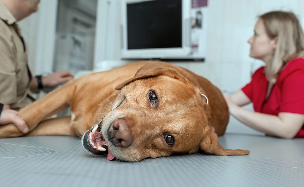
When a dog experiences abdominal pain, it can be distressing not only for the animal but also for the owner. The causes can range from minor gastrointestinal upset to serious, life-threatening conditions like organ torsion, internal bleeding, or tumors. Diagnosing the underlying issue accurately and swiftly is essential for appropriate treatment. In recent years, advanced imaging techniques such as Computed Tomography (CT) scans have become increasingly valuable tools in veterinary medicine, especially when dealing with abdominal discomfort.
Understanding Abdominal Pain in Dogs
Dogs cannot communicate their pain as humans do, so it’s crucial to recognize behavioral and physical signs that may indicate abdominal distress. Common symptoms include:
- Restlessness or inability to get comfortable
- Whimpering or vocalization
- Bloating or abdominal distension
- Vomiting or diarrhea
- Loss of appetite
- Guarding or sensitivity when the abdomen is touched
- Hunched posture or stretching repeatedly
These signs often prompt a visit to the veterinarian, who will start with a thorough physical examination and may proceed to blood tests, ultrasound, X-rays, or a CT scan to get to the root of the issue.
What Is a CT Scan?
A Computed Tomography (CT) scan is a non-invasive diagnostic imaging technique that uses X-ray technology to create detailed cross-sectional images of the body. Unlike traditional X-rays, which provide a flat image, CT scans produce a 3D representation of the internal organs, offering superior detail and contrast.
In veterinary medicine, CT scans are commonly used for:
- Investigating tumors or masses
- Detecting foreign bodies
- Diagnosing trauma or internal injuries
- Evaluating organ abnormalities
- Planning surgeries or treatment strategies
Why Use a CT Scan for Abdominal Pain?
The abdomen contains a complex array of organs, including the stomach, intestines, liver, spleen, kidneys, pancreas, bladder, and reproductive organs. Conditions affecting any of these structures can cause abdominal pain. CT scans are particularly useful because:
- High Resolution: CT imaging can reveal subtle lesions, abscesses, or masses that might not be visible on ultrasound or X-ray.
- 3D Imaging: This allows for better visualization of relationships between organs, which is critical in cases involving trauma, internal bleeding, or abnormal growths.
- Contrast Enhancement: CT can be performed with contrast agents to highlight blood vessels or differentiate between healthy and abnormal tissues.
- Speed: CT scans are relatively fast, often taking just a few minutes, which is essential when time is critical.
Preparing a Dog for a CT Scan
A CT scan requires the dog to remain completely still during imaging. Therefore, sedation or general anesthesia is typically administered to prevent movement and reduce stress. Prior to the scan:
- Fasting: The dog is usually fasted for several hours to prevent vomiting and reduce the risk of aspiration during anesthesia.
- Blood Work: Basic blood tests may be conducted to ensure the dog is healthy enough to undergo sedation.
- IV Access: An intravenous catheter is placed to administer anesthetics and contrast agents, if needed.
The CT Procedure
- Sedation/Anesthesia: Once the dog is safely sedated or anesthetized, it is positioned on the CT table.
- Imaging: The dog is passed through the CT scanner. A series of X-ray images are taken from multiple angles and processed by a computer to create cross-sectional views.
- Contrast Phase: In some cases, a contrast dye is injected into the bloodstream to enhance the images, especially useful in identifying vascular abnormalities or distinguishing cysts from solid masses.
- Monitoring: Throughout the procedure, the dog’s vital signs (heart rate, breathing, oxygen levels) are closely monitored by veterinary staff.
Interpreting the CT Scan
After the scan, a veterinary radiologist or specialist will analyze the images. This can help identify:
- Tumors or Cancer: CT can determine the size, shape, and spread of tumors more effectively than other imaging methods.
- Intestinal Obstructions: A CT can reveal blockages, twists (volvulus), or intussusceptions (telescoping of intestines).
- Organ Damage or Torsion: The spleen or stomach can twist (torsion), cutting off blood supply—CT is excellent at diagnosing these emergencies.
- Hernias: CT scans can clearly show abdominal contents pushing through a weakness in the abdominal wall.
- Internal Bleeding: Especially after trauma, CT is critical in identifying and locating hemorrhaging sources.
Benefits of CT Over Other Imaging Techniques
While ultrasound and X-rays are often first-line diagnostics for abdominal pain, CT offers distinct advantages:
- Greater Detail: Particularly useful in obese dogs or when the abdomen is filled with gas, which can obscure ultrasound images.
- Whole-Body Imaging: CT allows for evaluation of multiple organ systems at once, which is helpful when the source of pain is unclear.
- Surgical Planning: Surgeons rely on CT images to plan complex abdominal surgeries with greater accuracy and confidence.
Risks and Considerations
While CT scans are generally safe, there are a few considerations:
- Anesthesia Risks: As with any procedure requiring sedation, there are minor risks, especially in older or critically ill dogs.
- Radiation Exposure: Although low, there is some exposure to radiation, but the benefits far outweigh the risks in diagnostic cases.
- Cost: CT scans are more expensive than X-rays or ultrasounds, which may be a factor for some pet owners.
If your dog is experiencing unexplained abdominal discomfort, consult your veterinarian about whether a CT scan may be the right next step. Early diagnosis can make all the difference in ensuring your pet’s health and recovery.